THE SYSTEMIC CONSEQUENCES OF GOUT
Ada Kumar, MD, interviews Richard Johnson, MD, about the relationship between elevated uric acid levels and uncontrolled gout throughout the body. Dr Johnson answers crucial questions and discusses important research about the potential impact of systemic urate deposition that can lead to uncontrolled gout within the kidneys, vascular system, and other systems in the body.
Richard J. Johnson, MD, is the Tomas Berl Professor of Medicine in the Division of Renal Diseases and Hypertension at the University of Colorado Denver.
Ada Kumar, MD, is a board-certified radiologist and the former chief resident at Brigham and Women’s Hospital at Harvard Medical School.

Dr Richard Johnson,
Nephrologist
Nephrologist
A Webcast Series on Urate Acid Deposition and Gout With Dr Richard Johnson, Nephrologist
While they were once thought to be limited to bones and joints, uric acid crystals are now understood to also deposit in vital organs.1
In this webcast, Dr Johnson presents the case that elevated uric acid and gout should be evaluated for treatment by clinicians who treat patients for diseases such as chronic kidney disease (CKD).
Damage coming into focus: Inside Systemic Urate Deposition (SUD)
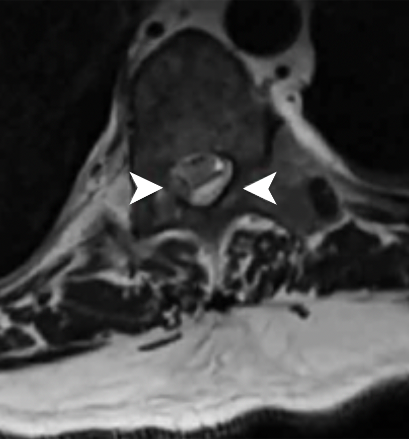
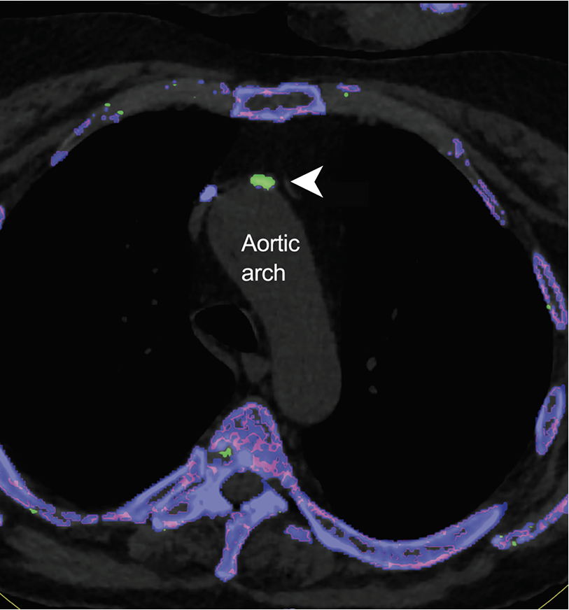
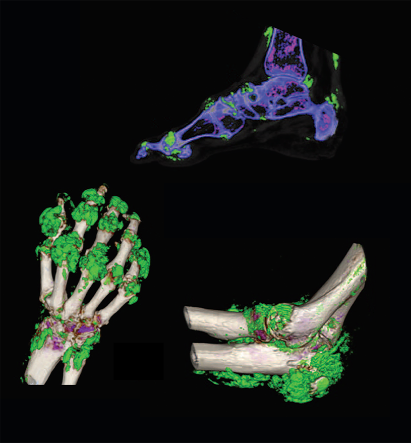
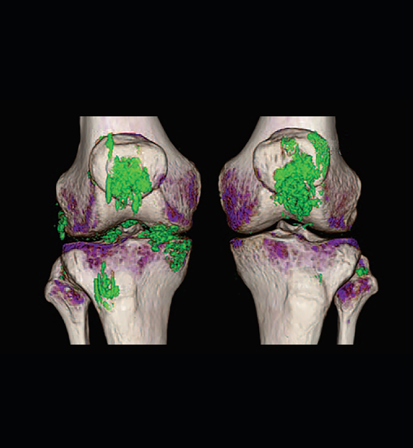
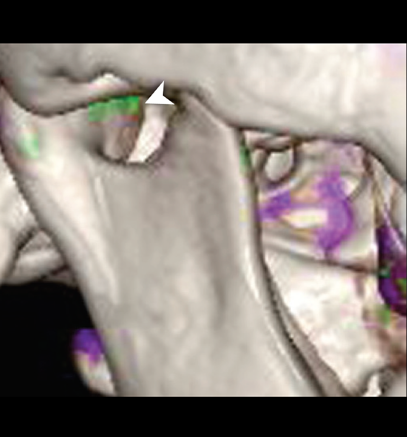
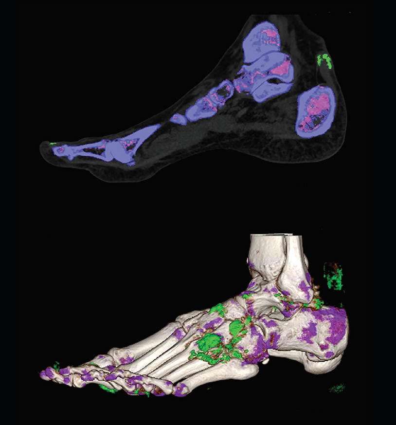
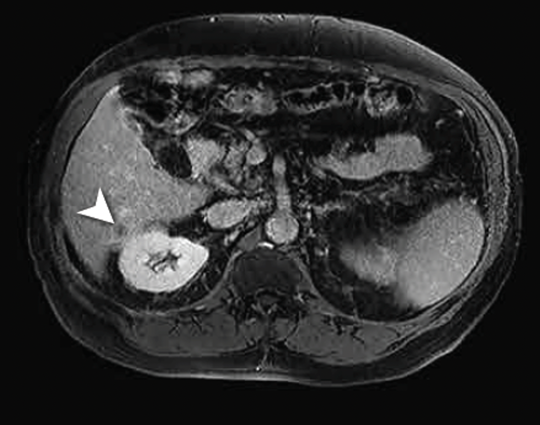
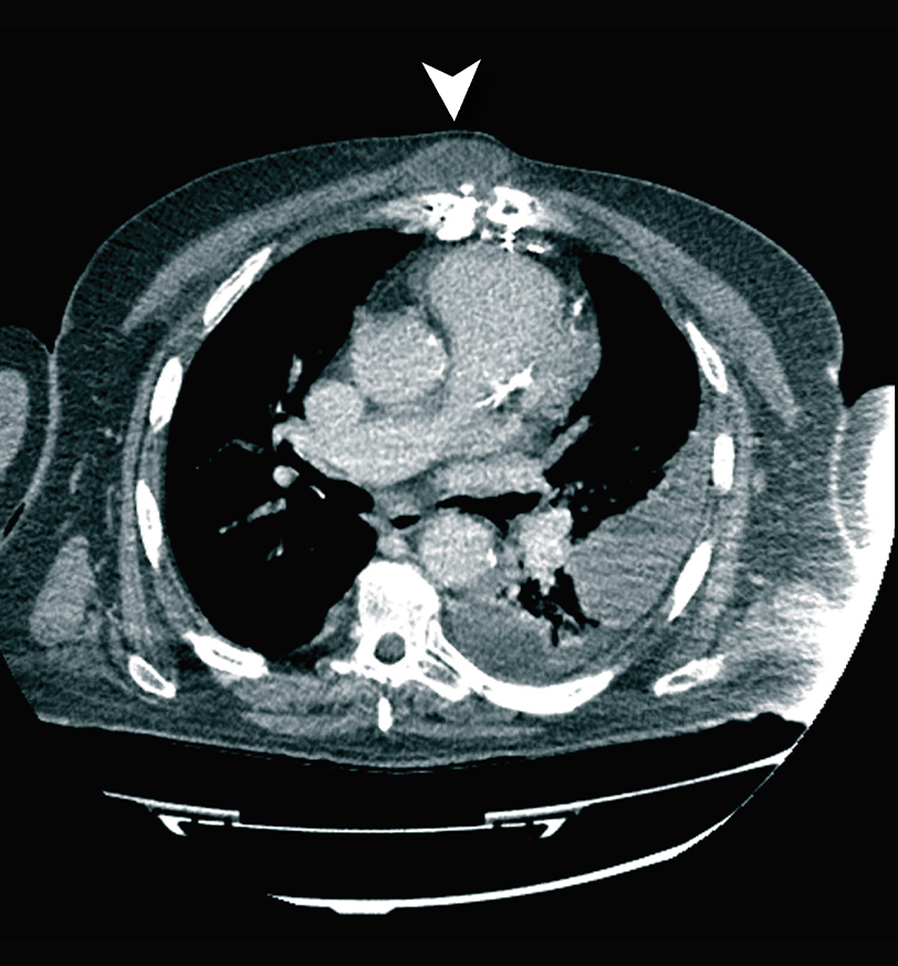
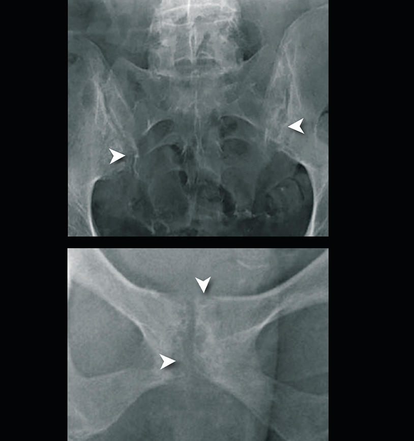
The extent of urate crystal deposition can be difficult to detect through physical examination alone2; thus, various imaging techniques can be employed to uncover systemic urate crystal deposition and its resulting damage.1,3
See systemic deposition now
Negatively birefringent crystal image (purple) used with permission from Park JJ, et al. BMJ Open. 2014;4(7):e005308.
Myocyte inclusions in the myocardium image used with permission from Frustaci A, et al. Ann Intern Med. 2020;172(5):363-365.
Kidney autopsy image used with permission from Nickeleit V, Mihatsh MJ. Nephrol Dial Transplant. 1997;12(9):1832-1838.
Spinal DECT scan used with permission from Wang JX, et al. BMC Rheumatol. 2020;4:22.
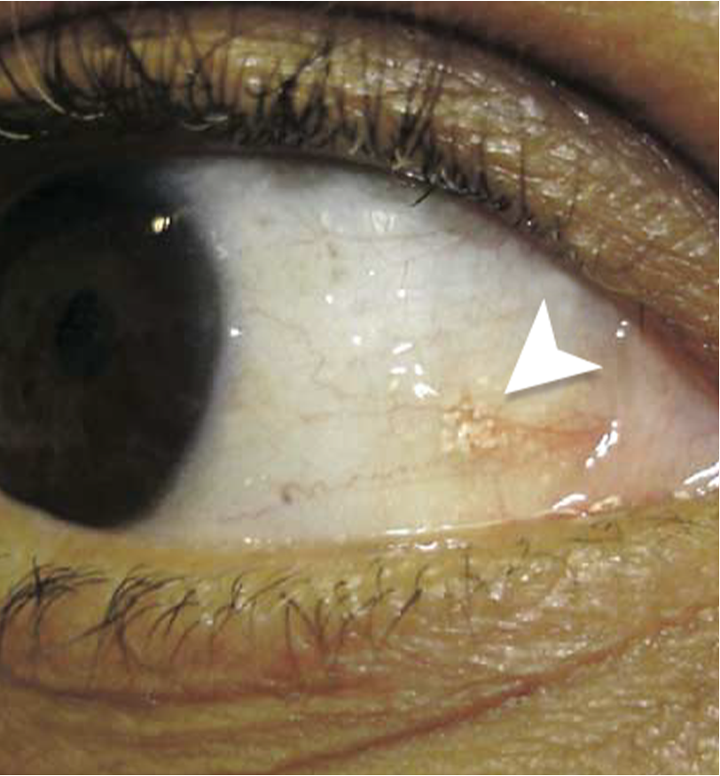
Fundoscopic photograph used with permission from Jiang Y, et al. BMC Ophthalmol. 2018;18(1):11.
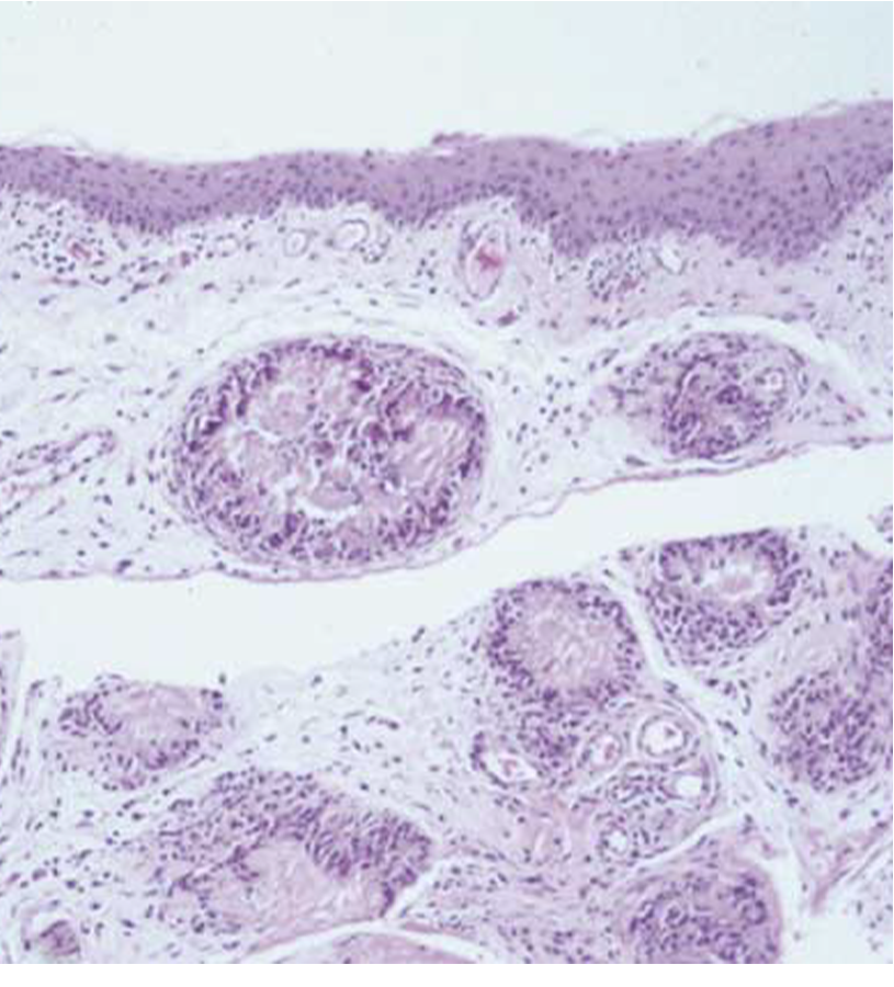
Skin image used with permission from Pattanaprichakul P, et al. Dermatol Pract Concept. 2014;4(4):33-35.
REFERENCES
-
Edwards NL. Primer on the Rheumatic Diseases. 13th ed. Springer;2008:241-262.
-
Choi HK, et al. Ann Rheum Dis. 2009;68:1609-1612.
-
Doghramji PP, et al. Postgrad Med. 2012;124:98-109.